Product Description
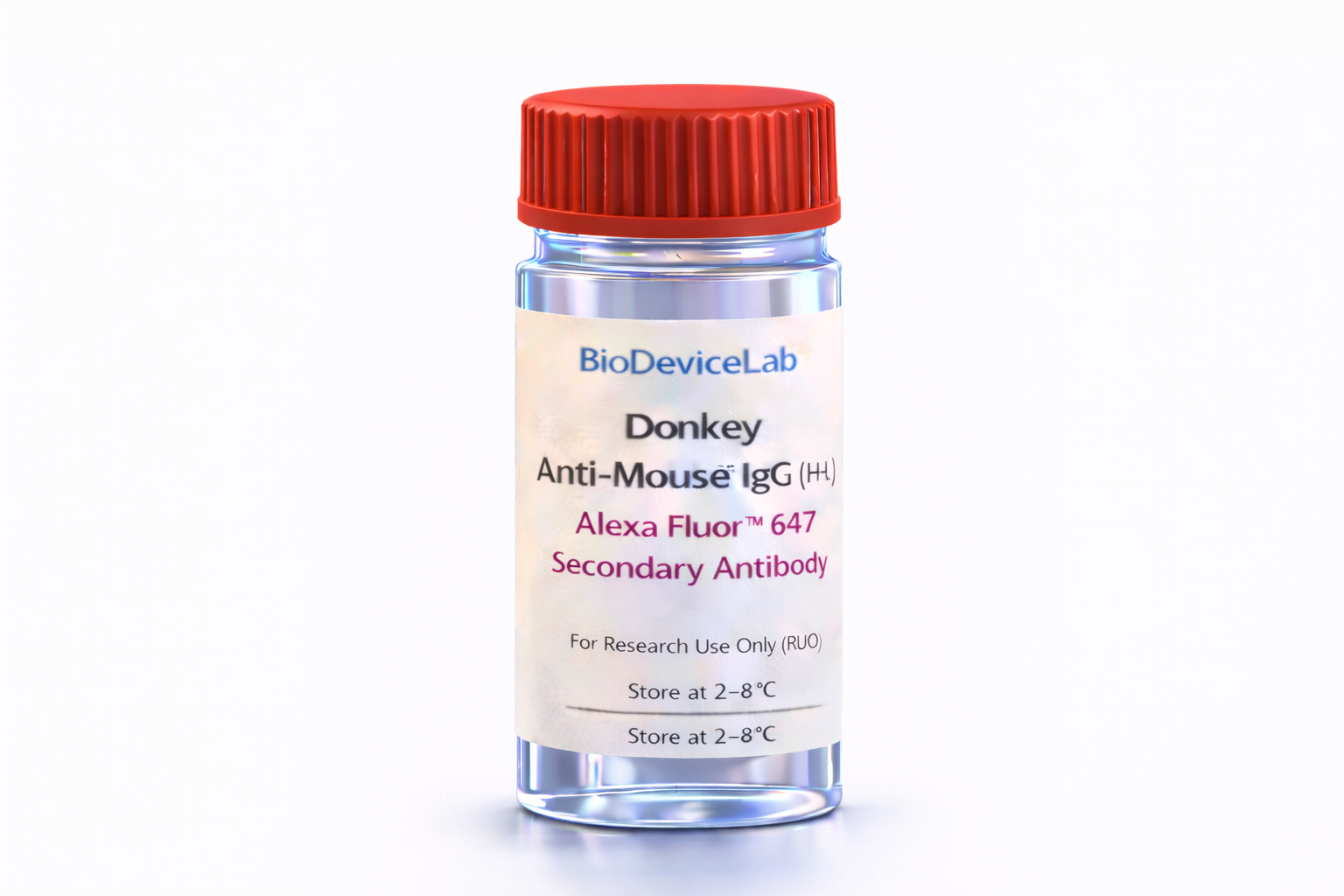
Donkey Anti-Mouse IgG (H+L) Alexa Fluor™ 647 is a purified secondary antibody that selectively binds to the heavy and light chains of mouse IgG primary antibodies. Conjugation to Alexa Fluor™ 647 enables bright, photostable fluorescence with minimal spectral overlap with green, yellow, and red fluorophores, supporting high-content and multiplexed imaging experiments.
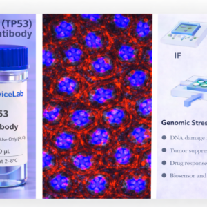
Alexa Fluor™ 647 exhibits an excitation maximum near 650 nm and an emission maximum near 668 nm, positioning it within the far-red region where cellular autofluorescence is low. This property improves signal-to-noise ratios in thick samples, tissues, organoids, and microfluidic or organ-on-chip platforms.
The antibody is suitable for indirect immunofluorescence workflows, where it amplifies signals from mouse primary antibodies while preserving spatial resolution and quantitative consistency. It is compatible with fixed cells, tissue sections, 3D cultures, and device-integrated staining protocols.
Intended Use
This product is intended for research applications including
• Immunofluorescence and immunocytochemistry
• Multiplex fluorescence imaging
• Confocal and super-resolution microscopy
• Organoid, spheroid, and 3D culture analysis
• Tissue section staining
• Biosensor and biodevice validation
• Cell phenotyping and marker colocalization studies
Principle of Operation
The antibody binds to mouse IgG primary antibodies via recognition of the Fc and Fab regions (H+L). Alexa Fluor™ 647 emits far-red fluorescence upon excitation, enabling
• Signal amplification of mouse primary antibodies
• Reduced background from cellular autofluorescence
• Spectral separation in multicolor imaging panels
• Quantitative and spatially resolved fluorescence detection
Technical Information
Target: Mouse IgG (H+L)
Host species: Donkey
Antibody type: Secondary antibody
Fluorophore: Alexa Fluor™ 647
Excitation maximum: ~650 nm
Emission maximum: ~668 nm
Isotype specificity: IgG heavy and light chains
Clonality: Polyclonal
Typical stock concentration (industry reference): ~0.5 mg/mL
Reactivity: Mouse IgG
Sample Volume and Typical Usage
Recommended volumes depend on assay format and platform.
Immunofluorescence / Immunocytochemistry
• Typical staining volume: 50–200 µL per sample
• Suitable for coverslips, chamber slides, well plates, tissue sections, and on-chip chambers
Organoid and 3D Culture Staining
• Typical staining volume: 100–500 µL per sample
• Volume depends on matrix density and construct size
Microfluidic and Biodevice Platforms
• Typical staining volume: 10–100 µL per device
• Adjust concentration for high surface-to-volume systems
General notes
• Optimize antibody dilution for each application
• Maintain consistent antibody-to-target ratios
• Protect fluorophore from prolonged light exposure
Selectivity and Compatibility
The antibody provides selective recognition of mouse IgG primary antibodies and is compatible with standard fixation buffers and blocking reagents. Alexa Fluor™ 647 is well suited for multiplex imaging with DAPI, Alexa Fluor™ 488, Alexa Fluor™ 555, and related fluorophores, enabling flexible panel design across imaging and device-based assays.
Package Contents
Each unit contains
• Purified BioDeviceLab Donkey Anti-Mouse IgG (H+L) Alexa Fluor™ 647 Secondary Antibody
• Lot-specific quality control documentation
Required Materials
• Mouse primary antibody
• Appropriate blocking and wash buffers
• Fluorescence microscope or imaging system with far-red detection
• Optional isotype and no-primary controls
Sample Handling and Use
• Store at 2–8 °C
• Do not freeze
• Minimize light exposure during storage and use
• Optimize dilution and incubation conditions per assay
Quality Control
Each lot is evaluated for binding specificity, fluorescence performance, and signal-to-background consistency to support reproducible imaging and quantitative analysis across platforms.
Applications
• Multiplex immunofluorescence imaging
• Tissue and organoid phenotyping
• Advanced microscopy workflows
• Biosensing and biodevice validation
• Cell-surface and intracellular marker detection