Product Description
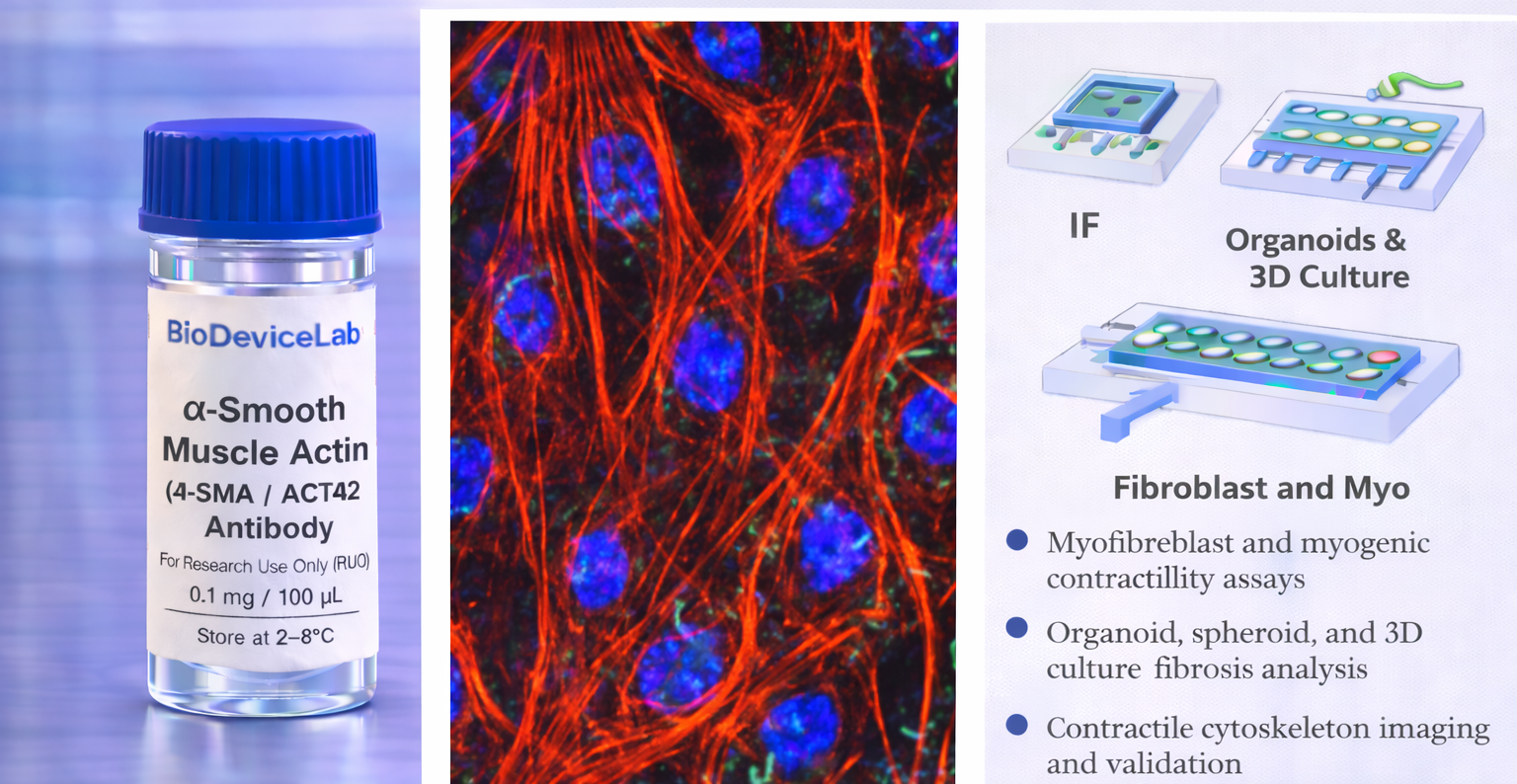
α-Smooth Muscle Actin, encoded by the ACTA2 gene, is a ~42 kDa actin isoform that is a core component of the contractile apparatus in smooth muscle cells and myofibroblasts. Unlike β-actin and γ-actin, which are ubiquitously expressed, α-SMA is selectively enriched in cells with contractile or force-generating functions.
Within cells, α-SMA assembles into prominent filament bundles that integrate with focal adhesions and stress fibers, enabling force transmission and mechanical remodeling of the extracellular environment. Upregulation of α-SMA is commonly associated with fibroblast activation, tissue remodeling, wound repair, and EMT-associated differentiation.
The BioDeviceLab α-SMA Antibody is supplied as a purified monoclonal antibody to support flexible assay design. It can be used with secondary antibodies for indirect detection, adapted for custom labeling strategies, or integrated into biosensor, biodevice, and high-content analytical workflows. The antibody is compatible with conventional culture systems as well as microfluidic, organ-on-chip, and advanced in vitro platforms.
Intended Use
This product is intended for research applications including
• Smooth muscle and myofibroblast identification
• Contractile cytoskeleton and force-generation studies
• Epithelial–mesenchymal transition and fibroblast activation analysis
• Immunofluorescence and immunocytochemistry imaging
• Organoid, spheroid, and 3D culture characterization
• Tissue engineering and regenerative medicine research
• Biosensor, biodevice, and assay biological validation
• Multiplex phenotyping of structural cell states
Principle of Operation
α-SMA is an intracellular actin filament protein. Binding of the antibody following permeabilization enables
• Visualization of contractile filament networks by immunofluorescence
• Identification of activated mesenchymal or myofibroblast phenotypes
• Assessment of cytoskeletal remodeling and mechanical adaptation
• Integration of contractility markers with adhesion or signaling readouts
Technical Information
Target antigen: α-Smooth Muscle Actin (α-SMA, ACTA2)
Alternative names: Smooth muscle α-actin
Protein type: Contractile actin isoform
Approximate molecular weight: ~42 kDa
Cellular localization: Cytoplasm (stress fibers and contractile filaments)
Clonality: Monoclonal
Host species: Mouse
Isotype: IgG (clone dependent)
Commonly used commercial clones: 1A4, ASM-1, EPR5368
Validated applications (clone dependent): IF/ICC, IHC, western blot, flow cytometry
Typical stock concentration (industry reference): approximately 0.5 mg/mL
Reactivity: Human (other species dependent on clone validation)
Sample Volume and Typical Usage
Recommended sample volumes depend on assay format and platform and should be optimized experimentally. Common working ranges are summarized below.
Immunofluorescence / Immunocytochemistry
• Typical staining volume: 50–200 µL per sample
• Applicable to coverslips, chamber slides, well plates, tissue sections, and on-chip chambers
• Permeabilization is required for intracellular filament access
Organoid and 3D Culture Staining
• Typical staining volume: 100–500 µL per sample
• Volume depends on matrix density and construct size
• Extended incubation times may be required for uniform penetration
Microfluidic and Biodevice Platforms
• Typical staining volume: 10–100 µL per device
• Volume depends on channel geometry and surface area
• Permeabilization conditions should be optimized for confined systems
General notes
• Antibody concentration is more critical than absolute volume
• Maintain consistent antibody-to-cell or antibody-to-surface ratios
• Optimization is recommended for each assay format and platform
Selectivity and Compatibility
α-SMA is selectively enriched in smooth muscle cells and activated mesenchymal or myofibroblast states and is typically low or absent in quiescent epithelial cells. The antibody is compatible with standard fixation and permeabilization protocols and performs reliably across conventional culture systems, 3D models, and microengineered platforms.
Package Contents
Each unit contains
• Purified BioDeviceLab α-SMA monoclonal antibody
• Lot-specific quality control documentation
Required Materials
• Fluorescent secondary antibody for unconjugated formats
• Fixation and permeabilization reagents
• Blocking and wash buffers
• Fluorescence microscope and/or flow cytometer
• Optional isotype and negative controls
Sample Handling and Use
• Store according to label instructions
• Avoid repeated freeze–thaw cycles
• Optimize staining conditions for each assay format
• Include appropriate controls for quantitative analyses
Quality Control
Each lot is evaluated for binding specificity and filamentous contractile staining patterns. The antibody is designed to support reproducible analysis of smooth muscle and myofibroblast phenotypes across diverse experimental systems.
Storage and Stability
• Store at 2–8 °C
• Do not freeze under standard storage conditions
• Protect from contamination and prolonged light exposure
• Proper storage preserves antibody stability and performance
Applications
• Smooth muscle and myofibroblast research
• Cytoskeletal contractility studies
• Tissue remodeling and regenerative medicine
• Organoid and 3D culture systems
• Immunofluorescence-based structural phenotyping
• Biosensor and biodevice validation
• Cell mechanics and force-generation studies