Product Description
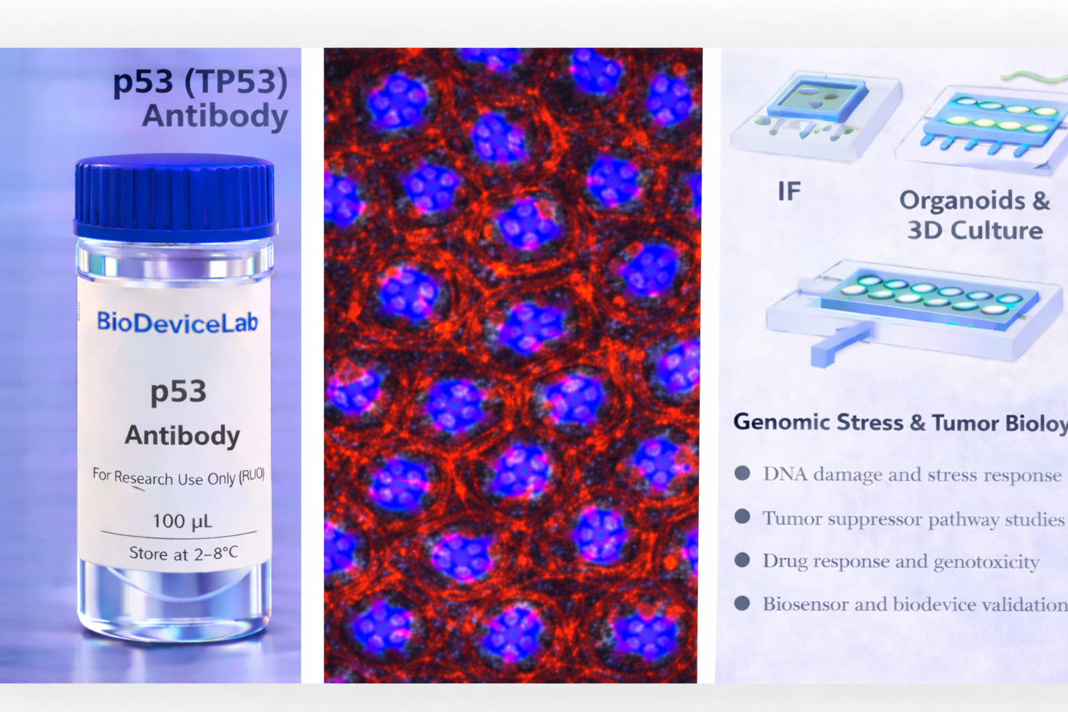
p53 (encoded by the TP53 gene) is a transcription factor that functions as a master regulator of cellular stress responses. Under normal conditions, p53 levels are kept low through rapid proteasomal degradation. In response to DNA damage, oncogenic stress, hypoxia, or metabolic perturbations, p53 becomes stabilized and activated, leading to transcriptional programs that control cell-cycle arrest, DNA repair, senescence, or apoptosis.
Mutations in TP53 are among the most common genetic alterations in human cancers, often resulting in loss of tumor suppressor function or accumulation of mutant p53 protein with altered transcriptional activity. As a result, p53 expression patterns, localization, and abundance are widely used to assess genomic integrity, oncogenic transformation, and therapeutic response.
Immunostaining of p53 typically reveals nuclear localization, reflecting its role as a transcriptional regulator. In stressed or transformed cells, increased nuclear accumulation of p53 can be readily detected and quantified. The BioDeviceLab p53 Antibody is supplied as a purified monoclonal antibody to support flexible assay design and integration into both conventional and advanced in vitro platforms.
The antibody is compatible with standard immunostaining workflows and is well suited for use in 2D cultures, 3D organoids, spheroids, and organ-on-chip systems where DNA damage, tumor suppressor pathways, or drug responses are investigated.
Intended Use
This product is intended for research applications including
• DNA damage and stress response analysis
• Tumor suppressor pathway studies
• Immunofluorescence and immunocytochemistry imaging
• Organoid, spheroid, and 3D culture characterization
• Organ-on-chip and microfluidic model validation
• Cancer biology and mutation-driven phenotyping
• Drug response and genotoxicity assessment
• Biosensor, biodevice, and assay biological validation
Principle of Operation
p53 is primarily localized in the nucleus following activation. After appropriate fixation and permeabilization, antibody binding enables
• Visualization of nuclear p53 accumulation by immunofluorescence
• Assessment of DNA damage–induced stress responses
• Identification of cells undergoing cell-cycle arrest or apoptosis
• Correlation of p53 activation with therapeutic or toxic stimuli
• Integration of genomic stress markers with proliferation or apoptosis readouts
Technical Information
Target antigen: p53 (TP53)
Alternative names: Tumor protein p53
Protein type: Transcription factor / tumor suppressor
Approximate molecular weight: ~53 kDa
Cellular localization: Predominantly nuclear
Clonality: Monoclonal
Host species: Mouse
Isotype: IgG (clone dependent)
Commonly used commercial clones: DO-1, DO-7, PAb240
Validated applications (clone dependent): IF/ICC, IHC, western blot
Typical stock concentration (industry reference): approximately 0.5 mg/mL
Reactivity: Human (other species dependent on clone validation)
Sample Volume and Typical Usage
Recommended sample volumes depend on assay format and platform and should be optimized experimentally. Common working ranges are summarized below.
Immunofluorescence / Immunocytochemistry
• Typical staining volume: 50–200 µL per sample
• Applicable to coverslips, chamber slides, well plates, tissue sections, and on-chip chambers
• Nuclear permeabilization is required
Organoid and 3D Culture Staining
• Typical staining volume: 100–500 µL per sample
• Volume depends on construct size and matrix density
• Extended incubation improves nuclear penetration
Microfluidic and Biodevice Platforms
• Typical staining volume: 10–100 µL per device
• Volume depends on channel geometry and chamber volume
• Flow-assisted staining may enhance uniform labeling
General notes
• Antibody concentration is more critical than absolute volume
• Maintain consistent antibody-to-cell ratios
• Optimization is recommended for each assay format
Selectivity and Compatibility
p53 is selectively enriched in cells undergoing stress, DNA damage, or oncogenic transformation. The antibody is compatible with commonly used fixation and staining protocols and performs reliably across conventional culture systems, 3D models, and microengineered platforms.
Package Contents
Each unit contains
• Purified BioDeviceLab p53 monoclonal antibody
• Lot-specific quality control documentation
Required Materials
• Fluorescent secondary antibody for unconjugated formats
• Fixation and permeabilization reagents
• Blocking and wash buffers
• Fluorescence microscope
• Optional isotype and negative controls
Sample Handling and Use
• Store according to label instructions
• Avoid repeated freeze–thaw cycles
• Optimize permeabilization for nuclear access
• Include appropriate controls for comparative analyses
Quality Control
Each lot is evaluated for binding specificity and nuclear staining performance. The antibody is designed to support reproducible analysis of tumor suppressor signaling across diverse experimental systems.
Storage and Stability
• Store at 2–8 °C
• Do not freeze under standard storage conditions
• Protect from contamination and prolonged light exposure
• Proper storage preserves antibody stability and performance
Applications
• Cancer biology and tumor suppressor research
• DNA damage and stress response studies
• Organoid and 3D culture systems
• Organ-on-chip and microfluidic models
• Drug screening and genotoxicity assays
• Immunofluorescence-based nuclear marker analysis
• Biosensor and biodevice validation