Product Description
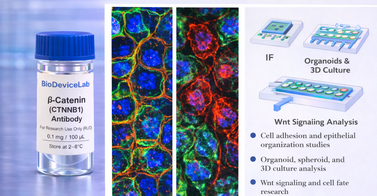
β-Catenin, encoded by the CTNNB1 gene, is a ~85–92 kDa protein that serves dual roles in epithelial biology: it links cadherins to the actin cytoskeleton at adherens junctions and acts as a transcriptional co-activator in the canonical Wnt signaling pathway. At the plasma membrane, β-catenin stabilizes cell–cell adhesion, while in the nucleus it regulates gene expression programs associated with proliferation, differentiation, and fate determination.
In resting cells, β-catenin is predominantly membrane-associated. Upon Wnt pathway activation or junctional remodeling, cytoplasmic accumulation and nuclear translocation occur. Immunostaining therefore yields context-dependent patterns—junctional, cytoplasmic, or nuclear—providing a sensitive readout of epithelial organization, signaling status, and phenotypic transitions.
The BioDeviceLab β-Catenin Antibody is supplied as a purified monoclonal antibody to support flexible assay design. It can be used with secondary antibodies for indirect detection, adapted for custom labeling strategies, or integrated into biosensor, biodevice, and high-content analytical workflows. The antibody is compatible with conventional culture systems as well as microfluidic, organ-on-chip, and advanced in vitro platforms.
Intended Use
This product is intended for research applications including
• Adherens junction visualization and epithelial organization studies
• Wnt/β-catenin signaling analysis
• Immunofluorescence and immunocytochemistry imaging
• Organoid, spheroid, and 3D culture characterization
• Cell fate, differentiation, and polarity studies
• Tissue engineering and regenerative medicine research
• Biosensor, biodevice, and assay biological validation
• Multiplex phenotyping and pathway analysis
Principle of Operation
β-Catenin (CTNNB1) localizes to adherens junctions and, upon signaling activation, to the cytoplasm and nucleus. Binding of the antibody following appropriate fixation/permeabilization enables
• Visualization of cell–cell adhesion complexes by immunofluorescence
• Detection of β-catenin redistribution associated with Wnt pathway activation
• Integration of structural and signaling readouts in multiplex assays
Technical Information
Target antigen: β-Catenin (CTNNB1)
Alternative names: Catenin beta-1
Protein type: Adherens junction protein and Wnt signaling effector
Approximate molecular weight: ~85–92 kDa
Cellular localization: Plasma membrane, cytoplasm, nucleus (context-dependent)
Clonality: Monoclonal
Host species: Mouse
Isotype: IgG (clone dependent)
Commonly used commercial clones: 14/β-Catenin, E-5, 15B8
Validated applications (clone dependent): IF/ICC, IHC, western blot, flow cytometry
Typical stock concentration (industry reference): approximately 0.5 mg/mL
Reactivity: Human (other species dependent on clone validation)
Sample Volume and Typical Usage
Recommended sample volumes depend on assay format and platform and should be optimized experimentally. Common working ranges are summarized below.
Immunofluorescence / Immunocytochemistry
• Typical staining volume: 50–200 µL per sample
• Applicable to coverslips, chamber slides, well plates, tissue sections, and on-chip chambers
• Permeabilization is required for cytoplasmic and nuclear detection
Organoid and 3D Culture Staining
• Typical staining volume: 100–500 µL per sample
• Volume depends on matrix density and construct size
• Extended incubation times may be required for uniform penetration
Microfluidic and Biodevice Platforms
• Typical staining volume: 10–100 µL per device
• Volume depends on channel geometry and surface area
• Fixation and permeabilization should be optimized for confined systems
General notes
• Antibody concentration is more critical than absolute volume
• Maintain consistent antibody-to-cell or antibody-to-surface ratios
• Optimization is recommended for each assay format and platform
Selectivity and Compatibility
β-Catenin provides a combined structural and signaling readout, enabling assessment of epithelial adhesion and pathway activation within the same system. The antibody is compatible with standard fixation and permeabilization protocols and performs reliably across conventional culture systems, 3D models, and microengineered platforms.
Package Contents
Each unit contains
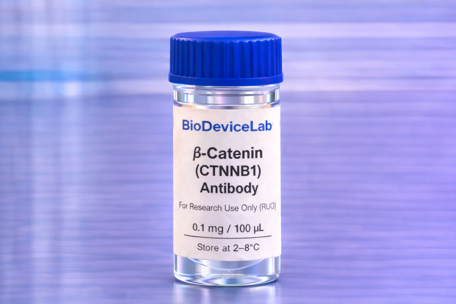
• Purified BioDeviceLab β-Catenin (CTNNB1) monoclonal antibody
• Lot-specific quality control documentation
Required Materials
• Fluorescent secondary antibody for unconjugated formats
• Fixation and permeabilization reagents
• Blocking and wash buffers
• Fluorescence microscope and/or flow cytometer
• Optional isotype and negative controls
Sample Handling and Use
• Store according to label instructions
• Avoid repeated freeze–thaw cycles
• Optimize staining conditions for each assay format
• Include appropriate controls for quantitative analyses
Quality Control
Each lot is evaluated for binding specificity and signal-to-background performance across membrane-associated and intracellular staining patterns. The antibody is designed to support reproducible adhesion and signaling analysis across diverse experimental systems.
Storage and Stability
• Store at 2–8 °C
• Do not freeze under standard storage conditions
• Protect from contamination and prolonged light exposure
• Proper storage preserves antibody stability and performance
Applications
• Adherens junction and epithelial organization studies
• Wnt/β-catenin signaling research
• Tissue engineering and regenerative medicine
• Organoid and 3D culture systems
• Immunofluorescence-based pathway analysis
• Biosensor and biodevice validation
• Cell fate and phenotypic transition studies