Product Description
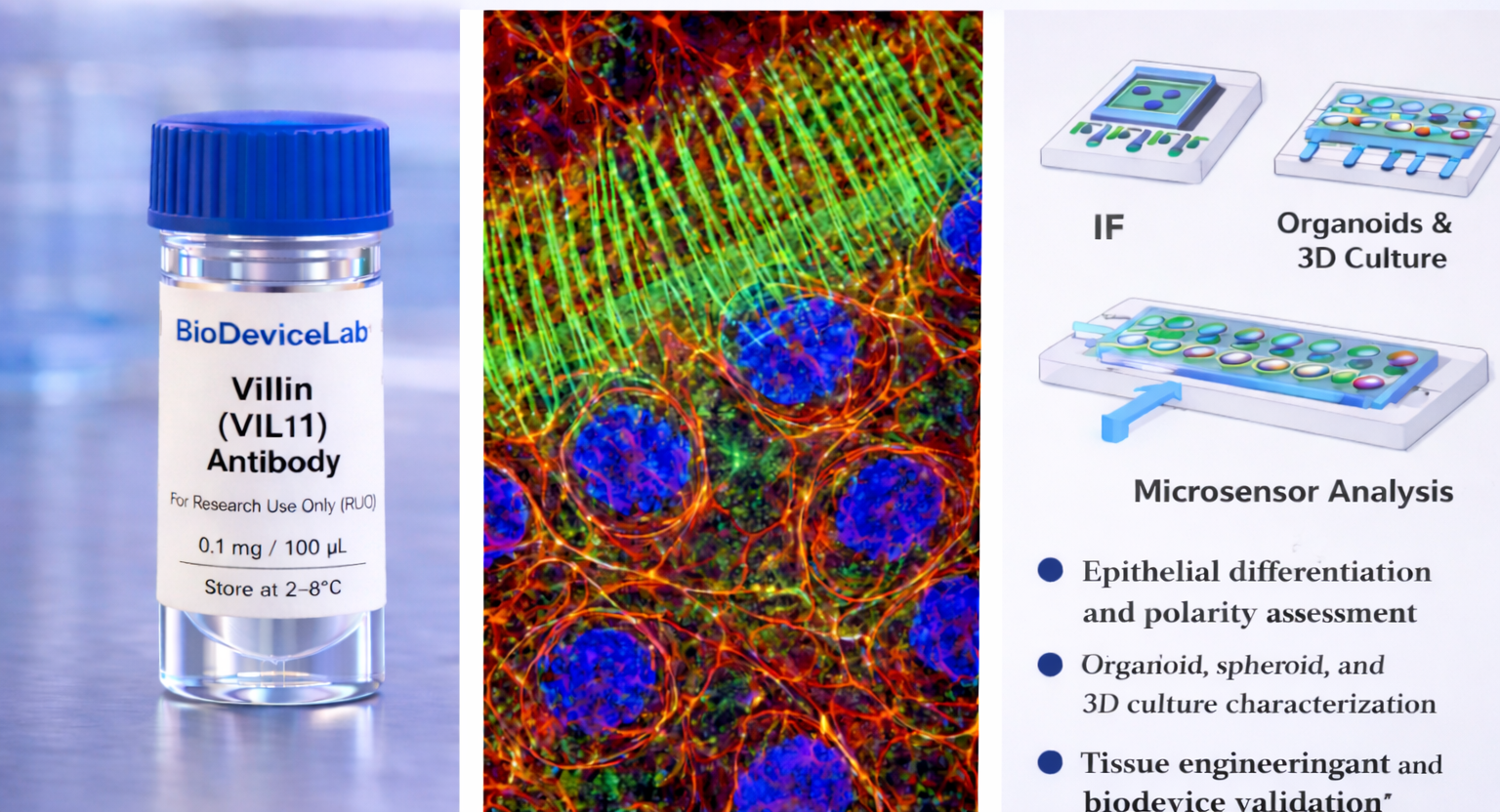
Villin, encoded by the VIL1 gene, is an actin-binding cytoskeletal protein with an approximate molecular weight of 95 kDa and is a key structural component of epithelial microvilli. Villin regulates actin filament bundling, severing, and reorganization in a calcium-dependent manner and plays a central role in epithelial cell morphology, differentiation, and apical surface specialization.
Villin is predominantly localized to the apical cytoplasm and microvillar structures of epithelial cells, where it contributes to brush border formation and maintenance. Immunostaining produces a characteristic apical and cytoskeletal pattern that provides a sensitive readout of epithelial differentiation state, polarization, and structural maturity.
The BioDeviceLab Villin Antibody is supplied as a purified monoclonal antibody to support flexible assay design. It is compatible with fixed 2D cultures, 3D tissue models, organoids, tissue sections, and microfluidic or biodevice-integrated platforms. Villin antibodies are widely used in epithelial biology, differentiation studies, tissue engineering, and translational biodevice validation.
Intended Use
This product is intended for research applications including
• Epithelial differentiation and polarity assessment
• Visualization of apical cytoskeletal organization
• Immunofluorescence and immunocytochemistry imaging
• Organoid, spheroid, and 3D culture characterization
• Cell identity analysis in mixed or co-culture systems
• Tissue engineering and regenerative medicine research
• Biosensor, biodevice, and assay biological validation
• Studies of epithelial maturation and structural remodeling
Principle of Operation
Villin (VIL1) is localized to the apical cytoplasm and microvilli of epithelial cells. Binding of the antibody following permeabilization enables
• Spatial visualization of epithelial apical organization by immunofluorescence
• Assessment of epithelial differentiation and polarization
• Integration into multiplex assays with junctional, membrane, or nuclear markers
Technical Information
Target antigen: Villin (VIL1)
Alternative names: Villin-1
Protein type: Actin-binding cytoskeletal protein
Approximate molecular weight: ~95 kDa
Cellular localization: Apical cytoplasm and microvilli
Clonality: Monoclonal
Host species: Mouse
Isotype: IgG (clone dependent)
Commonly used commercial clones: ID2C3, 1D2C3, CWWB1
Validated applications (clone dependent): IF/ICC, immunohistochemistry, western blot
Typical stock concentration (industry reference): approximately 0.5 mg/mL
Reactivity: Human (other species dependent on clone validation)
Sample Volume and Typical Usage
Recommended sample volumes depend on assay format and platform and should be optimized experimentally. Common working ranges are summarized below.
Immunofluorescence / Immunocytochemistry
• Typical staining volume: 50–200 µL per sample
• Applicable to coverslips, chamber slides, well plates, tissue sections, and on-chip staining chambers
• Samples must be permeabilized to enable intracellular access
Organoid and 3D Culture Staining
• Typical staining volume: 100–500 µL per sample
• Volume depends on matrix composition, organoid density, and vessel geometry
• Extended incubation times may be required to ensure apical labeling
Microfluidic and Biodevice Platforms
• Typical staining volume: 10–100 µL per device
• Volume depends on channel dimensions and surface area
• Permeabilization conditions should be optimized to preserve cytoskeletal architecture
General notes
• Antibody concentration is more critical than absolute volume
• Maintain consistent antibody-to-surface ratios
• Optimization is recommended for each assay format and platform
Selectivity and Compatibility
Villin is selectively enriched in differentiated epithelial cells and provides a robust marker of apical organization and epithelial maturation. The antibody is compatible with standard fixation and permeabilization protocols and performs reliably in conventional culture systems, tissue sections, and microfluidic or biodevice-integrated workflows.
Package Contents
Each unit contains
• Purified BioDeviceLab Villin (VIL1) monoclonal antibody
• Lot-specific quality control documentation
Required Materials
• Fluorescent secondary antibody for unconjugated formats
• Fixation and permeabilization buffers
• Blocking and wash buffers compatible with IF
• Fluorescence microscope
• Optional isotype and negative controls
Sample Handling and Use
• Store according to label instructions
• Avoid repeated freeze–thaw cycles
• Optimize antibody concentration, volume, and permeabilization conditions
• Use appropriate controls for comparative analysis
Quality Control
Each lot is evaluated for binding specificity and apical cytoskeletal staining performance. The antibody is designed to support reproducible epithelial differentiation profiling across diverse experimental platforms and longitudinal studies.
Storage and Stability
• Store at 2–8 °C
• Do not freeze under standard storage conditions
• Protect from contamination and prolonged light exposure
• Proper storage preserves antibody stability and performance
Applications
• Epithelial differentiation and polarity biology
• Tissue engineering and regenerative medicine
• Organoid and 3D culture systems
• Immunofluorescence-based cytoskeletal analysis
• Biosensing and biodevice validation
• Cell identity and maturation studies