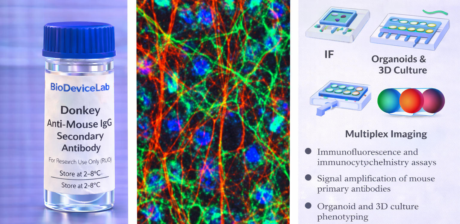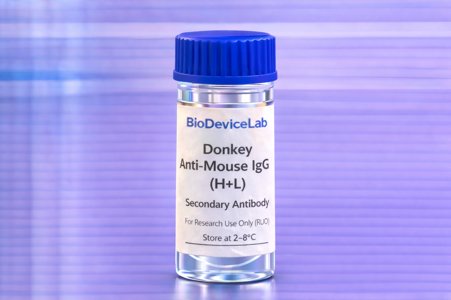
Product Description
Donkey anti-mouse IgG (H+L) secondary antibodies are widely used for indirect detection of mouse primary antibodies in imaging-based assays. This antibody recognizes both the heavy and light chains of mouse IgG, enabling robust signal amplification while maintaining compatibility with a broad range of mouse monoclonal primary antibodies.
To minimize non-specific binding and cross-species reactivity, the BioDeviceLab Donkey Anti-Mouse IgG (H+L) Secondary Antibody is highly cross-adsorbed against immunoglobulins from multiple species. This feature is particularly important for complex samples, co-culture systems, tissue sections, and microfluidic or organ-on-chip platforms where background signal must be tightly controlled.
The antibody is supplied in an unconjugated or fluorophore-conjugated format (depending on configuration) to support flexible assay design. It is compatible with standard fixation and staining protocols and performs reliably in conventional culture systems as well as advanced bioengineered environments.
Intended Use
This product is intended for research applications including
• Immunofluorescence and immunocytochemistry detection of mouse primary antibodies
• Signal amplification in indirect antibody staining workflows
• Multiplex imaging assays using spectrally distinct fluorophores
• Organoid, spheroid, and 3D culture staining
• Tissue section and whole-mount imaging
• Biosensor, biodevice, and organ-on-chip validation
• Cell phenotyping and marker visualization studies
Principle of Operation
The secondary antibody binds to the Fc and light chain regions of mouse IgG primary antibodies. Upon binding, it enables
• Fluorescent or chromogenic signal amplification
• Spatial visualization of target antigens labeled by mouse primaries
• Integration of indirect detection into multiplex staining panels
Technical Information
Target specificity: Mouse IgG (H+L)
Antibody type: Secondary antibody
Host species: Donkey
Cross-adsorption: Highly cross-adsorbed against multiple species
Clonality: Polyclonal
Isotype: IgG
Conjugation: Unconjugated or fluorophore-conjugated (format dependent)
Validated applications: IF/ICC, IHC (clone and conjugate dependent)
Reactivity: Mouse IgG
Sample Volume and Typical Usage
Recommended sample volumes depend on assay format and platform and should be optimized experimentally. Common working ranges are summarized below.
Immunofluorescence / Immunocytochemistry
• Typical staining volume: 50–200 µL per sample
• Applicable to coverslips, chamber slides, well plates, tissue sections, and on-chip chambers
• Typical dilution range: 1:200–1:1000 (optimize per assay)
Organoid and 3D Culture Staining
• Typical staining volume: 100–500 µL per sample
• Volume depends on construct size and matrix density
• Extended incubation may improve penetration
Microfluidic and Biodevice Platforms
• Typical staining volume: 10–100 µL per device
• Volume depends on channel geometry and surface area
• Lower concentrations may reduce background in confined systems
General notes
• Optimize dilution to balance signal intensity and background
• Use consistent secondary antibody conditions across experiments
• Protect fluorophore-conjugated antibodies from light
Selectivity and Compatibility
The antibody selectively binds mouse IgG and shows minimal cross-reactivity with immunoglobulins from other species due to extensive cross-adsorption. It is compatible with commonly used blocking buffers, fixation reagents, and staining protocols and performs reliably across conventional and microengineered platforms.
Package Contents
Each unit contains
• Purified BioDeviceLab Donkey Anti-Mouse IgG (H+L) Secondary Antibody
• Lot-specific quality control documentation
Required Materials
• Mouse primary antibody
• Blocking and wash buffers
• Fluorescence or brightfield microscope (depending on detection method)
• Optional nuclear or cytoskeletal counterstains
Sample Handling and Use
• Store according to label instructions
• Avoid repeated freeze–thaw cycles
• Protect from prolonged light exposure if fluorophore-conjugated
• Optimize dilution and incubation time for each assay
Quality Control
Each lot is evaluated for specificity, signal intensity, and background performance in indirect detection assays. The antibody is designed to support reproducible and high-contrast imaging across diverse experimental systems.
Storage and Stability
• Store at 2–8 °C
• Do not freeze under standard storage conditions
• Protect from contamination and light exposure
• Proper storage preserves antibody stability and performance
Applications
• Indirect immunofluorescence and immunocytochemistry
• Multiplex imaging assays
• Organoid and 3D culture staining
• Tissue imaging and histological studies
• Biosensor and biodevice validation
• Cell phenotyping and marker visualization