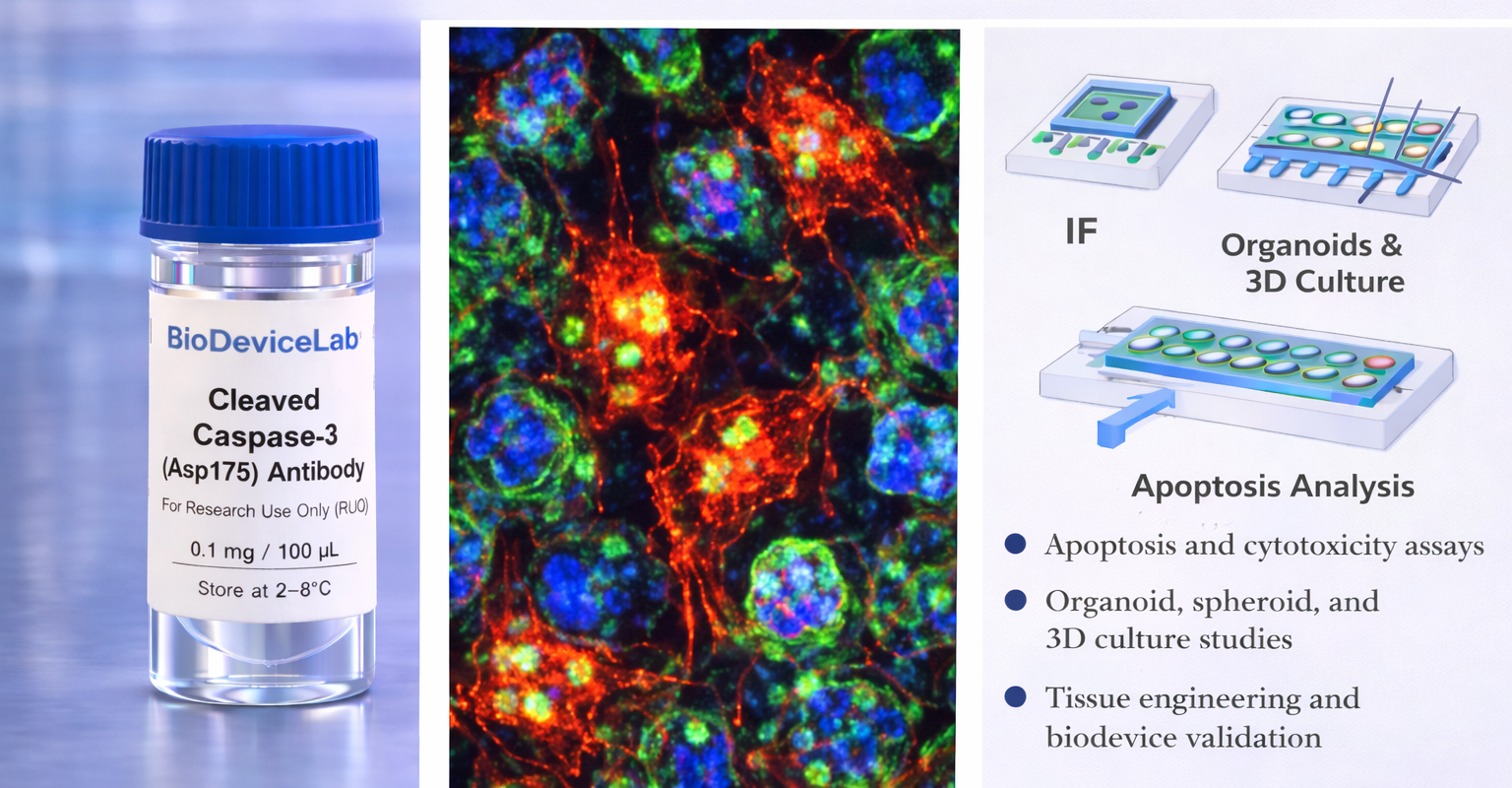
Product Description
Caspase-3 is a cysteine protease that plays a central role in the execution phase of apoptosis. During apoptotic signaling, inactive pro-caspase-3 is proteolytically cleaved into its active form, commonly referred to as cleaved Caspase-3. This activated enzyme mediates the cleavage of multiple cellular substrates, leading to DNA fragmentation, cytoskeletal reorganization, and irreversible commitment to cell death.
Cleaved Caspase-3 is localized primarily in the cytoplasm and nucleus of apoptotic cells and is absent in viable or early-stage cells. Antibody staining produces a characteristic intracellular pattern that enables sensitive and specific identification of cells undergoing apoptosis. Because cleavage represents an irreversible activation step, cleaved Caspase-3 is considered one of the most reliable markers of late-stage programmed cell death.
The BioDeviceLab Cleaved Caspase-3 Antibody is supplied as a purified monoclonal antibody to support flexible assay design. It can be used with secondary antibodies for indirect detection, adapted for custom labeling strategies, or integrated into biosensor, biodevice, and high-content analytical workflows. The antibody is compatible with conventional culture systems as well as microfluidic, organ-on-chip, and advanced in vitro platforms.
Intended Use
This product is intended for research applications including
• Detection and quantification of apoptotic cells
• Programmed cell death and cytotoxicity studies
• Immunofluorescence and immunocytochemistry imaging
• Organoid, spheroid, and 3D culture studies
• Tissue engineering and regenerative medicine research
• Drug response and toxicity assessment
• Biosensor, biodevice, and assay biological validation
• Multiplex cell fate and phenotyping assays
Principle of Operation
Cleaved Caspase-3 is generated exclusively during execution-phase apoptosis. Binding of the antibody following permeabilization enables
• Identification of apoptotic cells by immunofluorescence
• Quantitative assessment of apoptosis levels within populations
• Spatial mapping of cell death in 2D, 3D, and device-based systems
• Integration of apoptosis metrics with proliferation or structural markers
Technical Information
Target antigen: Cleaved Caspase-3 (Asp175)
Alternative names: Active Caspase-3, CPP32
Protein type: Apoptosis executioner protease (cleaved form)
Approximate molecular weight: ~17–19 kDa (cleaved fragments)
Cellular localization: Cytoplasm and nucleus (apoptotic cells)
Clonality: Monoclonal
Host species: Rabbit or mouse (clone dependent)
Isotype: IgG (clone dependent)
Commonly used commercial clones: Asp175, C92-605, 5A1E
Validated applications (clone dependent): IF/ICC, IHC, western blot, flow cytometry
Typical stock concentration (industry reference): approximately 0.5 mg/mL
Reactivity: Human (other species dependent on clone validation)
Sample Volume and Typical Usage
Recommended sample volumes depend on assay format and platform and should be optimized experimentally. Common working ranges are summarized below.
Immunofluorescence / Immunocytochemistry
• Typical staining volume: 50–200 µL per sample
• Applicable to coverslips, chamber slides, well plates, tissue sections, and on-chip chambers
• Permeabilization is required for intracellular access
Flow Cytometry
• Typical staining volume: 50–100 µL per test
• Typical cell input: approximately 1 × 10⁵ to 1 × 10⁶ cells per test
• Fixation and permeabilization are required prior to staining
Organoid and 3D Culture Staining
• Typical staining volume: 100–500 µL per sample
• Volume depends on matrix density and construct size
• Extended incubation times may be required for antibody penetration
Microfluidic and Biodevice Platforms
• Typical staining volume: 10–100 µL per device
• Volume depends on channel geometry and surface area
• Permeabilization conditions should be optimized for confined systems
General notes
• Antibody concentration is more critical than absolute volume
• Maintain consistent antibody-to-cell ratios
• Optimization is recommended for each assay format and platform
Selectivity and Compatibility
Cleaved Caspase-3 is selectively present in apoptotic cells and is absent in viable or non-apoptotic populations, enabling clear discrimination of programmed cell death. The antibody is compatible with standard fixation, permeabilization, and staining protocols and performs reliably across conventional culture systems, 3D models, and microengineered platforms.
Package Contents
Each unit contains
• Purified BioDeviceLab Cleaved Caspase-3 monoclonal antibody
• Lot-specific quality control documentation
Required Materials
• Fluorescent secondary antibody for unconjugated formats
• Fixation and permeabilization reagents
• Blocking and wash buffers
• Fluorescence microscope and/or flow cytometer
• Optional isotype and negative controls
Sample Handling and Use
• Store according to label instructions
• Avoid repeated freeze–thaw cycles
• Optimize staining conditions for each assay format
• Include appropriate controls for quantitative analyses
Quality Control
Each lot is evaluated for binding specificity and signal-to-background performance. The antibody is designed to support reproducible apoptosis analysis across diverse experimental systems and longitudinal studies.
Storage and Stability
• Store at 2–8 °C
• Do not freeze under standard storage conditions
• Protect from contamination and prolonged light exposure
• Proper storage preserves antibody stability and performance
Applications
• Apoptosis and cell death analysis
• Drug toxicity and treatment response studies
• Cancer and translational research
• Tissue engineering and regenerative medicine
• Organoid and 3D culture systems
• Immunofluorescence-based apoptosis assays
• Biosensor and biodevice validation
• Cell fate and phenotyping studies