Product Description
The BioDeviceLab 100 nm Gold Nanoparticles are monodisperse colloidal particles designed for applications requiring a pronounced optical signature and robust surface area. At ~100 nm diameter, these nanoparticles exhibit a significant localized surface plasmon resonance (LSPR) and enhanced light scattering, distinguishing them from smaller gold nanoparticle formats in optical and colorimetric assays.
Stabilized with citrate/tannic acid ligands in an aqueous formulation, 100 nm gold nanoparticles offer excellent colloidal stability and compatibility with biological and engineering workflows, including biosensor calibration, lateral flow assay design, microfluidic integration, and surface functionalization studies.
The broader LSPR and scatter-dominated optical profile of 100 nm nanoparticles enable strong UV–Vis absorbance and light scatter signatures, facilitating sensitive detection in plasmonic biosensing and enhanced contrast in microscopy and analytical platforms.
Intended Use
This product is intended for research applications including:
• Optical and plasmonic biosensor development
• Light scattering and enhanced colorimetric assays
• Passive conjugation of proteins, antibodies, and ligands
• Microfluidic and lab-on-chip integration
• Imaging and analytical contrast enhancement
• Biodevice calibration and benchmarking
Assay Principle & Optical Behavior
Gold nanoparticles exhibit localized surface plasmon resonance (LSPR) — collective oscillation of conduction electrons in response to incident light.
• The LSPR peak and light scattering intensity are influenced by particle size and environment.
• Larger particles (such as 100 nm) produce strong light scattering, making them effective for optical and lateral-flow readouts.
• Binding events or changes in local refractive index modulate optical signals, enabling sensing.
• High surface area at this size supports effective passive adsorption for conjugation.
Optical & Physical Characteristics
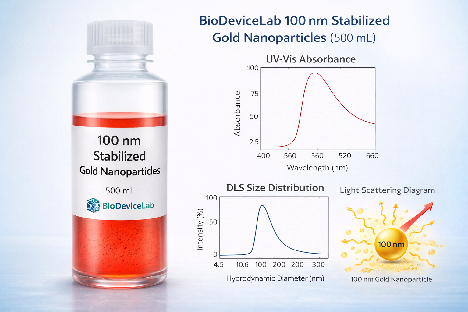
Nominal particle diameter: ~100 nm
Characteristic optical features: Strong absorbance and light scattering
Peak SPR wavelength: ~525–540 nm (size-dependent)
Particle concentration: ~2.5 × 10¹¹ particles/mL
Weight concentration: ~2.5 mg/mL
Molar extinction coefficient: ~1.0 × 10⁹ M⁻¹·cm⁻¹
Size dispersity: <15%
Surface area per particle: ~3.14 × 10⁴ nm²
Surface-to-volume ratio: ~0.2
Particle mass: ~3.27 × 10⁻¹⁴ g
Molar mass: ~4.90 × 10⁹ g/mol
Molar concentration: ~5.1 × 10⁻¹¹ M
Supplied volume: 500 mL
Selectivity & Compatibility
Gold nanoparticles are chemically inert and are broadly compatible with biological molecules, assays, and device interfaces. They can be integrated with proteins, nucleic acids, polymers, and optical/electrochemical interfaces without interfering with recognition events.
Package Contents
Each unit contains:
• 500 mL of 100 nm citrate-stabilized gold nanoparticle colloidal suspension
• Optical characterization and quality control documentation
Required Equipment (Not Provided)
• UV–Vis spectrophotometer
• Dynamic Light Scattering (DLS) instrument (recommended)
• Standard laboratory glassware and micropipettes
• Aqueous buffers appropriate for conjugation or downstream workflows
Sample Handling & Use
• Gently swirl before use to ensure uniform suspension.
• Avoid vigorous shaking or sonication that may induce aggregation.
• Dilute with compatible aqueous buffers for downstream applications.
• Minimize repeated contact or contamination of the stock solution.
Quality Control
• Each production lot is verified for optical features, particle concentration, and size distribution.
• Batch consistency supports reproducibility in biosensor development and biodevice workflows.
• Optical and size metrics follow standardized QC practices.
Storage & Stability
• Store at 4–25 °C.
• Do not freeze.
• Protect from prolonged direct light exposure.
• Proper storage maintains colloidal stability and performance throughout the product’s shelf life.
Limitations
• For research use only.
• Not intended for diagnostic or therapeutic use.
• Performance may vary with buffer conditions and functionalization strategies.
Applications
• Optical and plasmonic biosensor development
• Light-scattering and enhanced colorimetric assays
• Protein and antibody conjugate preparation
• Microfluidic and lab-on-chip systems
• Imaging contrast and analytical probing
• Biodevice calibration and benchmarking


