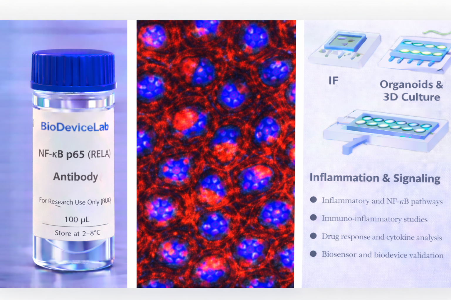
Product Description
NF-κB p65, also known as RELA, is a major subunit of the NF-κB transcription factor complex. In resting cells, p65 is retained in the cytoplasm through interaction with inhibitory IκB proteins. Upon activation by inflammatory cytokines, microbial products, oxidative stress, or mechanical cues, IκB proteins are degraded, allowing p65 to translocate into the nucleus where it regulates the expression of a broad range of genes involved in inflammation, immunity, cell survival, and stress responses.
NF-κB signaling plays a central role in epithelial barrier function, immune cell activation, wound repair, and host–microbe interactions. Dysregulated or sustained NF-κB activation is implicated in chronic inflammation, fibrosis, autoimmune disorders, and cancer progression. As a result, p65 localization and activation state are widely used as readouts of inflammatory signaling and cellular stress.
Immunostaining of NF-κB p65 enables visualization of cytoplasmic versus nuclear localization, providing a direct and spatially resolved measure of pathway activation. The BioDeviceLab NF-κB p65 Antibody is supplied as a purified monoclonal antibody to support flexible assay design and integration into both conventional and advanced in vitro platforms.
The antibody is compatible with standard immunostaining workflows and is well suited for use in 2D cultures, 3D organoids, spheroids, and organ-on-chip systems where inflammatory signaling and immune–epithelial interactions are investigated.
Intended Use
This product is intended for research applications including
• Inflammatory signaling and NF-κB pathway analysis
• Immunofluorescence and immunocytochemistry imaging
• Organoid, spheroid, and 3D culture characterization
• Organ-on-chip and microfluidic model validation
• Immune–epithelial interaction studies
• Stress and cytokine response analysis
• Drug screening targeting inflammatory pathways
• Biosensor, biodevice, and assay biological validation
Principle of Operation
NF-κB p65 undergoes cytoplasmic-to-nuclear translocation upon pathway activation. Following fixation and permeabilization, antibody binding enables
• Visualization of NF-κB nuclear translocation by immunofluorescence
• Assessment of inflammatory pathway activation
• Quantification of signaling responses to cytokines or stressors
• Correlation of inflammatory signaling with epithelial or immune markers
• Integration of pathway activation with functional phenotypes
Technical Information
Target antigen: NF-κB p65 (RELA)
Alternative names: RELA, NF-κB subunit p65
Protein type: Transcription factor
Approximate molecular weight: ~65 kDa
Cellular localization: Cytoplasmic and nuclear (activation dependent)
Clonality: Monoclonal
Host species: Mouse
Isotype: IgG (clone dependent)
Commonly used commercial clones: F-6, D14E12, L8F6
Validated applications (clone dependent): IF/ICC, IHC, western blot
Typical stock concentration (industry reference): approximately 0.5 mg/mL
Reactivity: Human (other species dependent on clone validation)
Sample Volume and Typical Usage
Recommended sample volumes depend on assay format and platform and should be optimized experimentally. Common working ranges are summarized below.
Immunofluorescence / Immunocytochemistry
• Typical staining volume: 50–200 µL per sample
• Applicable to coverslips, chamber slides, well plates, tissue sections, and on-chip chambers
• Nuclear permeabilization is required to assess translocation
Organoid and 3D Culture Staining
• Typical staining volume: 100–500 µL per sample
• Volume depends on construct size and matrix density
• Extended incubation improves nuclear penetration
Microfluidic and Biodevice Platforms
• Typical staining volume: 10–100 µL per device
• Volume depends on channel geometry and chamber volume
• Flow-assisted staining may enhance uniform labeling
General notes
• Antibody concentration is more critical than absolute volume
• Maintain consistent antibody-to-cell ratios
• Optimization is recommended for each assay format
Selectivity and Compatibility
NF-κB p65 is selectively activated and translocated to the nucleus in response to inflammatory and stress signals. The antibody is compatible with commonly used fixation and staining protocols and performs reliably across conventional culture systems, 3D models, and microengineered platforms.
Package Contents
Each unit contains
• Purified BioDeviceLab NF-κB p65 monoclonal antibody
• Lot-specific quality control documentation
Required Materials
• Fluorescent secondary antibody for unconjugated formats
• Fixation and permeabilization reagents
• Blocking and wash buffers
• Fluorescence microscope
• Optional isotype and negative controls
Sample Handling and Use
• Store according to label instructions
• Avoid repeated freeze–thaw cycles
• Optimize permeabilization for nuclear access
• Include appropriate controls for comparative analyses
Quality Control
Each lot is evaluated for binding specificity and cytoplasmic-to-nuclear translocation staining performance. The antibody is designed to support reproducible analysis of inflammatory signaling across diverse experimental systems.
Storage and Stability
• Store at 2–8 °C
• Do not freeze under standard storage conditions
• Protect from contamination and prolonged light exposure
• Proper storage preserves antibody stability and performance
Applications
• Inflammation and immune signaling research
• Organoid and 3D culture systems
• Organ-on-chip and microfluidic models
• Cytokine and stress-response studies
• Drug screening targeting NF-κB signaling
• Immunofluorescence-based pathway activation analysis
• Biosensor and biodevice validation


