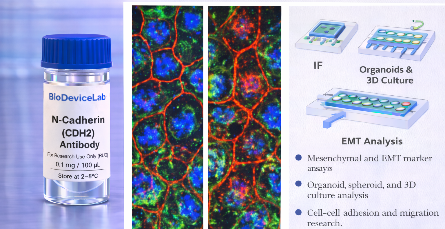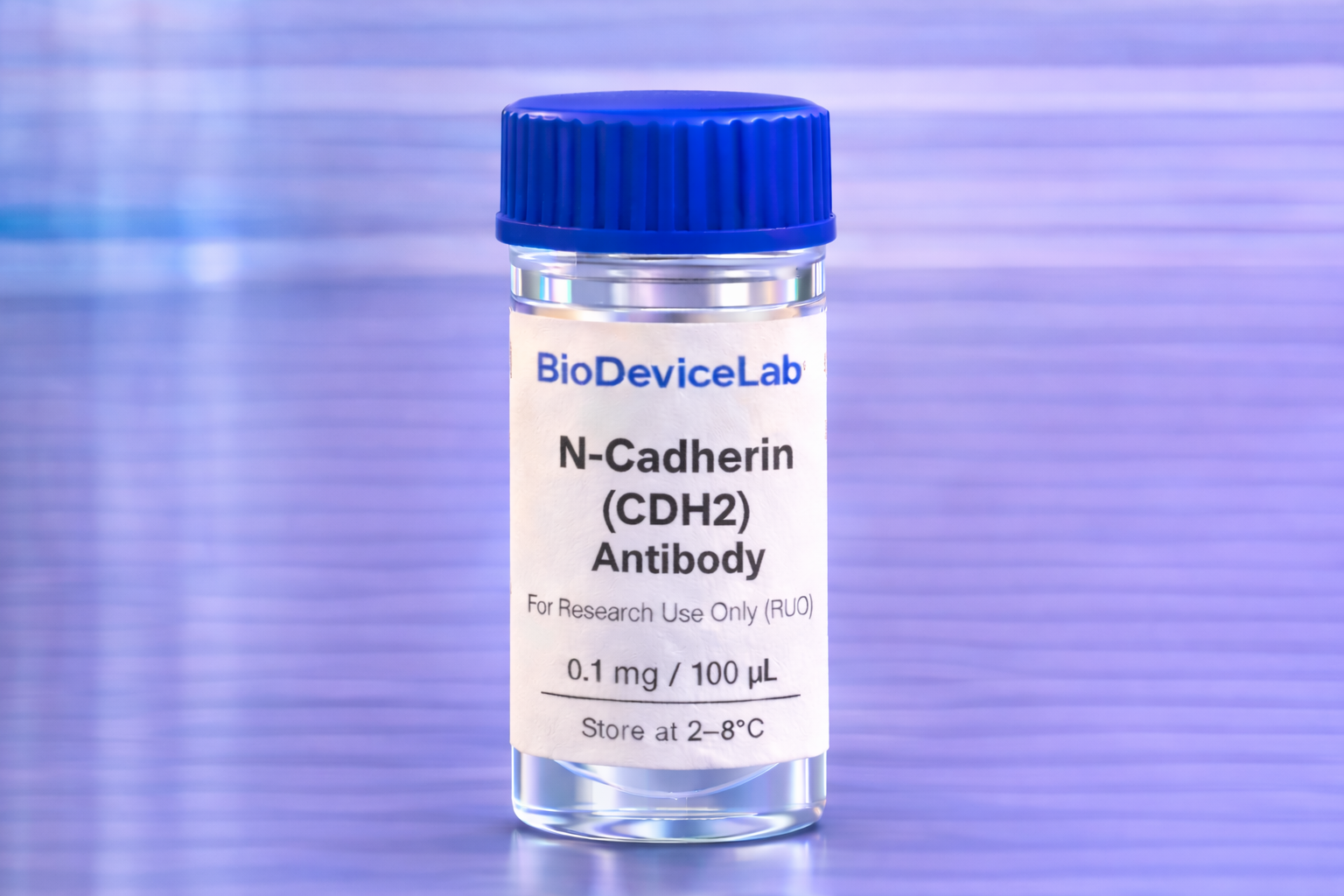
Product Description
N-Cadherin, encoded by the CDH2 gene, is a ~130–135 kDa transmembrane glycoprotein belonging to the classical cadherin family. It mediates homophilic cell–cell adhesion through calcium-dependent interactions and plays a central role in tissue morphogenesis, cell migration, and mechanical coupling between cells.
Unlike E-cadherin, which is characteristic of stable epithelial junctions, N-cadherin is predominantly expressed in mesenchymal, neural, and migratory cell populations. Upregulation of N-cadherin often accompanies epithelial–mesenchymal transition and is associated with increased cellular motility, plasticity, and altered tissue organization.
The BioDeviceLab N-Cadherin Antibody is supplied as a purified monoclonal antibody to support flexible assay design. It can be used with secondary antibodies for indirect detection, adapted for custom labeling strategies, or integrated into biosensor, biodevice, and high-content analytical workflows. The antibody is compatible with conventional culture systems as well as microfluidic, organ-on-chip, and advanced in vitro platforms.
Intended Use
This product is intended for research applications including
• Mesenchymal cell identification and characterization
• Epithelial–mesenchymal transition (EMT) analysis
• Cell–cell adhesion and junctional remodeling studies
• Immunofluorescence and immunocytochemistry imaging
• Organoid, spheroid, and 3D culture characterization
• Tissue engineering and regenerative medicine research
• Biosensor, biodevice, and assay biological validation
• Multiplex phenotyping and cell state analysis
Principle of Operation
N-Cadherin is localized to the plasma membrane at cell–cell junctions. Binding of the antibody following appropriate fixation enables
• Visualization of mesenchymal and migratory junctional structures
• Assessment of cadherin switching during EMT
• Correlation of adhesion remodeling with cytoskeletal or signaling markers
• Integration of structural readouts into multiplex assays
Technical Information
Target antigen: N-Cadherin (CDH2)
Alternative names: Cadherin-2, NCAD
Protein type: Calcium-dependent cell adhesion glycoprotein
Approximate molecular weight: ~130–135 kDa
Cellular localization: Plasma membrane, adherens junctions
Clonality: Monoclonal
Host species: Mouse
Isotype: IgG (clone dependent)
Commonly used commercial clones: 32/N-Cadherin, 13A9, 6G11
Validated applications (clone dependent): IF/ICC, IHC, western blot, flow cytometry
Typical stock concentration (industry reference): approximately 0.5 mg/mL
Reactivity: Human (other species dependent on clone validation)
Sample Volume and Typical Usage
Recommended sample volumes depend on assay format and platform and should be optimized experimentally. Common working ranges are summarized below.
Immunofluorescence / Immunocytochemistry
• Typical staining volume: 50–200 µL per sample
• Applicable to coverslips, chamber slides, well plates, tissue sections, and on-chip chambers
• Membrane staining may require mild permeabilization depending on fixation
Organoid and 3D Culture Staining
• Typical staining volume: 100–500 µL per sample
• Volume depends on matrix density and construct size
• Extended incubation times may be required for uniform penetration
Microfluidic and Biodevice Platforms
• Typical staining volume: 10–100 µL per device
• Volume depends on channel geometry and surface area
• Staining conditions should be optimized for confined systems
General notes
• Antibody concentration is more critical than absolute volume
• Maintain consistent antibody-to-cell or antibody-to-surface ratios
• Optimization is recommended for each assay format and platform
Selectivity and Compatibility
N-Cadherin is selectively enriched in mesenchymal and EMT-associated cell populations and is typically reduced in stable epithelial states. The antibody is compatible with standard fixation and staining protocols and performs reliably across conventional culture systems, 3D models, and microengineered platforms.
Package Contents
Each unit contains
• Purified BioDeviceLab N-Cadherin (CDH2) monoclonal antibody
• Lot-specific quality control documentation
Required Materials
• Fluorescent secondary antibody for unconjugated formats
• Fixation and permeabilization reagents
• Blocking and wash buffers
• Fluorescence microscope and/or flow cytometer
• Optional isotype and negative controls
Sample Handling and Use
• Store according to label instructions
• Avoid repeated freeze–thaw cycles
• Optimize staining conditions for each assay format
• Include appropriate controls for quantitative analyses
Quality Control
Each lot is evaluated for binding specificity and membrane-localized staining patterns. The antibody is designed to support reproducible EMT and adhesion profiling across diverse experimental systems.
Storage and Stability
• Store at 2–8 °C
• Do not freeze under standard storage conditions
• Protect from contamination and prolonged light exposure
• Proper storage preserves antibody stability and performance
Applications
• EMT and cadherin-switching studies
• Mesenchymal phenotype analysis
• Tissue engineering and regenerative medicine
• Organoid and 3D culture systems
• Immunofluorescence-based adhesion studies
• Biosensor and biodevice validation
• Cell migration and plasticity research