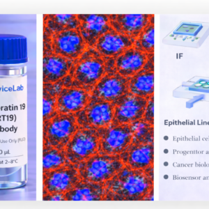
Product Description
Histone H2AX is a variant of the H2A histone family that becomes rapidly phosphorylated at serine 139 in response to DNA double-strand breaks. This phosphorylated form, known as γH2AX, is one of the earliest and most sensitive indicators of DNA damage. Upon damage induction, γH2AX forms distinct nuclear foci surrounding break sites, serving as a platform for the recruitment of DNA repair proteins and checkpoint signaling complexes.
γH2AX plays a central role in coordinating DNA damage signaling, chromatin remodeling, and repair pathway activation. Its formation is triggered by ionizing radiation, oxidative stress, replication stress, chemotherapeutic agents, and mechanical or microenvironmental perturbations. Quantification of γH2AX foci is widely used to assess genomic instability, DNA repair capacity, and treatment-induced genotoxicity.
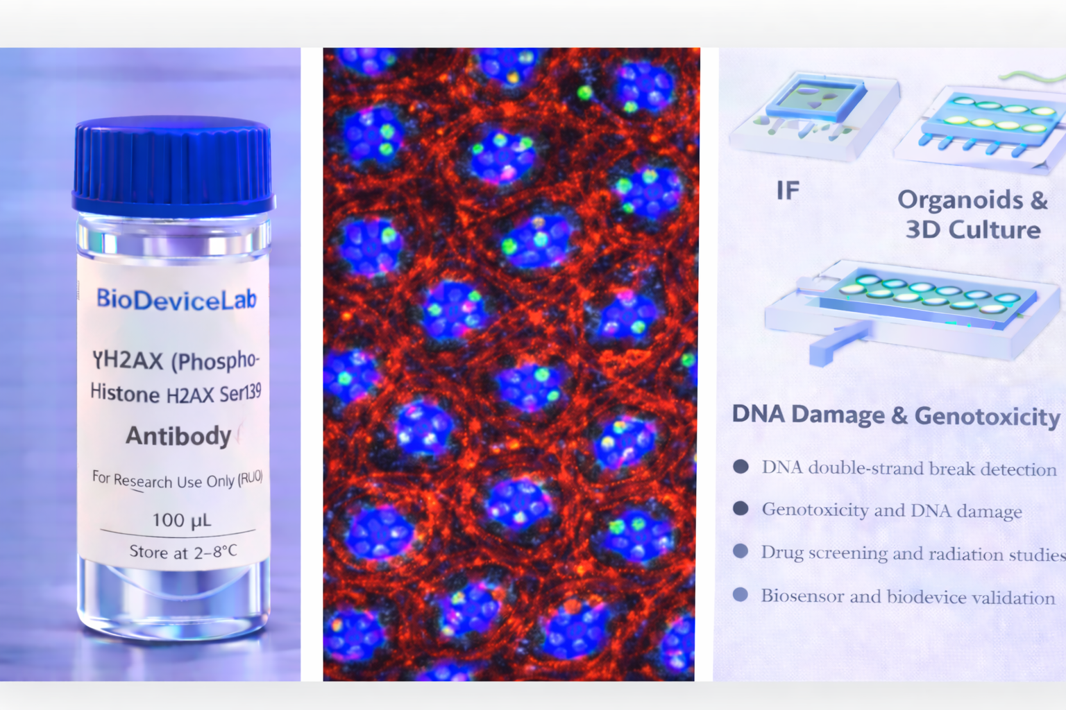
Immunostaining of γH2AX reveals discrete nuclear foci whose number, size, and intensity correlate with the extent of DNA damage. This makes γH2AX particularly well suited for quantitative imaging, high-content screening, and time-resolved damage-repair studies. The BioDeviceLab γH2AX Antibody is supplied as a purified monoclonal antibody to support flexible assay design and integration into both conventional and advanced in vitro platforms.
The antibody is compatible with standard immunostaining workflows and is well suited for use in 2D cultures, 3D organoids, spheroids, and organ-on-chip systems where DNA damage, replication stress, or treatment response must be evaluated.
Intended Use
This product is intended for research applications including
• DNA double-strand break detection
• Genotoxicity and DNA damage assessment
• Immunofluorescence and immunocytochemistry imaging
• Organoid, spheroid, and 3D culture characterization
• Organ-on-chip and microfluidic model validation
• Drug screening and radiation response studies
• DNA repair pathway and checkpoint signaling analysis
• Biosensor, biodevice, and assay biological validation
Principle of Operation
γH2AX is localized in the nucleus at sites of DNA double-strand breaks. Following fixation and permeabilization, antibody binding enables
• Visualization of DNA damage foci by immunofluorescence
• Quantification of DNA damage burden at the single-cell level
• Monitoring of damage induction and repair kinetics
• Correlation of genomic stress with proliferation or apoptosis markers
• Integration of DNA damage readouts with signaling and phenotypic data
Technical Information
Target antigen: Phospho-Histone H2AX (Ser139)
Alternative names: γH2AX
Protein type: Phosphorylated histone variant
Approximate molecular weight: ~15 kDa
Cellular localization: Nuclear foci
Clonality: Monoclonal
Host species: Mouse
Isotype: IgG (clone dependent)
Commonly used commercial clones: JBW301, 2F3, EP854(2)Y
Validated applications (clone dependent): IF/ICC, IHC, western blot
Typical stock concentration (industry reference): approximately 0.5 mg/mL
Reactivity: Human (other species dependent on clone validation)
Sample Volume and Typical Usage
Recommended sample volumes depend on assay format and platform and should be optimized experimentally. Common working ranges are summarized below.
Immunofluorescence / Immunocytochemistry
• Typical staining volume: 50–200 µL per sample
• Applicable to coverslips, chamber slides, well plates, tissue sections, and on-chip chambers
• Nuclear permeabilization is required
Organoid and 3D Culture Staining
• Typical staining volume: 100–500 µL per sample
• Volume depends on construct size and matrix density
• Extended incubation improves nuclear penetration
Microfluidic and Biodevice Platforms
• Typical staining volume: 10–100 µL per device
• Volume depends on channel geometry and chamber volume
• Flow-assisted staining may enhance uniform labeling
General notes
• Antibody concentration is more critical than absolute volume
• Maintain consistent antibody-to-cell ratios
• Optimization is recommended for each assay format
Selectivity and Compatibility
γH2AX is selectively enriched at sites of DNA double-strand breaks and is absent in undamaged chromatin. The antibody is compatible with commonly used fixation and staining protocols and performs reliably across conventional culture systems, 3D models, and microengineered platforms.
Package Contents
Each unit contains
• Purified BioDeviceLab γH2AX monoclonal antibody
• Lot-specific quality control documentation
Required Materials
• Fluorescent secondary antibody for unconjugated formats
• Fixation and permeabilization reagents
• Blocking and wash buffers
• Fluorescence microscope
• Optional isotype and negative controls
Sample Handling and Use
• Store according to label instructions
• Avoid repeated freeze–thaw cycles
• Optimize permeabilization for nuclear access
• Include appropriate controls for quantitative analyses
Quality Control
Each lot is evaluated for binding specificity and discrete nuclear foci staining performance. The antibody is designed to support reproducible quantification of DNA damage across diverse experimental systems.
Storage and Stability
• Store at 2–8 °C
• Do not freeze under standard storage conditions
• Protect from contamination and prolonged light exposure
• Proper storage preserves antibody stability and performance
Applications
• DNA damage and repair research
• Genotoxicity and radiation biology
• Organoid and 3D culture systems
• Organ-on-chip and microfluidic models
• Drug screening and treatment response studies
• Immunofluorescence-based nuclear foci analysis
• Biosensor and biodevice validation