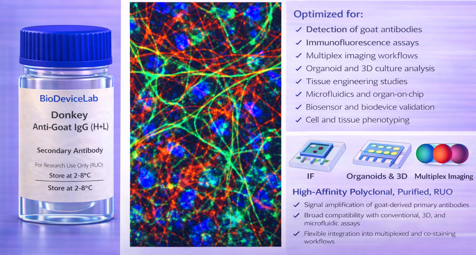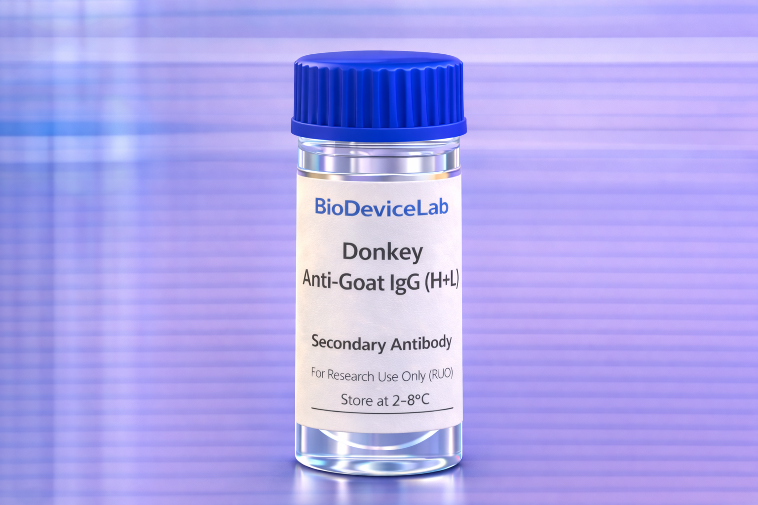
Product Description
The BioDeviceLab Donkey Anti-Goat IgG (H+L) Secondary Antibody is a purified polyclonal antibody raised in donkey and directed against both the heavy and light chains of goat immunoglobulin G. By recognizing the conserved regions of goat IgG, this secondary antibody enables reliable detection and amplification of signals generated by goat primary antibodies across a wide range of biological targets.
Secondary antibodies play a central role in immunoassays by amplifying primary antibody signals, improving sensitivity, and enabling flexible assay design through indirect labeling strategies. This antibody is suitable for use with fluorescent, enzymatic, or custom-conjugated detection systems and is compatible with standard fixation and staining protocols.
The antibody is formulated to perform consistently in traditional cell culture assays as well as advanced applications including organoids, 3D cultures, tissue engineering constructs, and microfluidic or organ-on-chip platforms. Its broad compatibility makes it well suited for imaging-based studies, multiplexed assays, and biodevice validation workflows.
Intended Use
This product is intended for research applications including
• Detection of goat primary antibodies
• Immunofluorescence and immunocytochemistry assays
• Multiplex imaging and co-staining workflows
• Organoid, spheroid, and 3D culture analysis
• Tissue engineering and regenerative medicine research
• Microfluidic and organ-on-chip staining
• Biosensor and biodevice biological validation
• Cell and tissue phenotyping studies
Principle of Operation
The Donkey Anti-Goat IgG (H+L) Secondary Antibody binds to the heavy and light chains of goat IgG molecules. This interaction enables
• Amplification of signals from goat primary antibodies
• Indirect detection using fluorescent or enzymatic labels
• Flexible integration into multiplexed and multi-marker assays
Technical Information
Target antigen: Goat IgG (H+L)
Host species: Donkey
Clonality: Polyclonal
Isotype: IgG
Specificity: Goat IgG heavy and light chains
Cross-adsorption: Application-dependent
Format: Purified, unconjugated
Typical stock concentration (industry reference): ~1 mg/mL
Reactivity: Goat primary antibodies
Sample Volume and Typical Usage
Recommended usage should be optimized for each assay format. Common working ranges include
Immunofluorescence / Immunocytochemistry
• Typical staining volume: 50–200 µL per sample
• Suitable for coverslips, chamber slides, multiwell plates, and on-chip staining
Flow Cytometry (when applicable)
• Typical staining volume: 50–100 µL per test
• Antibody concentration should be titrated per assay
Organoids and 3D Cultures
• Typical staining volume: 100–500 µL per sample
• Longer incubation may be required for dense matrices
Microfluidic and Biodevice Platforms
• Typical staining volume: 10–100 µL per device
• Concentration and flow conditions should be optimized
General notes
• Optimize dilution and incubation time for each application
• Maintain consistent antibody-to-sample ratios
• Include appropriate controls in multiplex assays
Selectivity and Compatibility
This secondary antibody provides broad compatibility with goat primary antibodies and is suitable for use in complex biological systems. It performs reliably in standard laboratory assays as well as advanced imaging, 3D culture, and biodevice-integrated workflows.
Package Contents
Each unit contains
• Purified BioDeviceLab Donkey Anti-Goat IgG (H+L) Secondary Antibody
• Lot-specific quality control documentation
Required Materials
• Goat-derived primary antibody
• Appropriate blocking and wash buffers
• Fluorescence microscope or imaging system
• Optional isotype and negative controls
Sample Handling and Use
• Store according to label instructions
• Avoid repeated freeze–thaw cycles
• Optimize dilution, volume, and incubation conditions
• Protect from contamination and prolonged light exposure
Quality Control
Each lot is evaluated for binding performance and signal consistency to support reproducible detection across imaging, cytometry, and device-based assays.
Storage and Stability
• Store at 2–8 °C
• Do not freeze under standard storage conditions
• Proper storage preserves antibody integrity and performance
Applications
• Immunofluorescence and immunocytochemistry
• Multiplex imaging workflows
• Organoid and 3D culture characterization
• Tissue engineering and regenerative medicine
• Biosensor and biodevice validation
• Cell and tissue phenotyping